Blood Vessels Labeled / How The Heart Blood Vessels Work Heart Vascular Institute Temple Health : The common cartoid artery extends from the brachiocephalic artery.
byAdmin-
0
Blood Vessels Labeled / How The Heart Blood Vessels Work Heart Vascular Institute Temple Health : The common cartoid artery extends from the brachiocephalic artery.. Disclosure • the material and the illustrations are adopted from the textbook human anatomy and physiology / ninth edition/ The microvasculature is composed of blood vessels that are smaller than 100 microns may only be seen through the microscope. The iliac, femoral, popliteal and tibial (calf) veins are the deep veins in the legs. Liver anatomy blood supply 19 photos of the liver anatomy blood supply anatomical location of liver, blood vessels that carry blood to the liver, dual blood supply to liver, functional anatomy of liver, liver and its functions, liver on the human body, normal anatomy of the liver, position of liver, inner body, anatomical location of … Blood vessels are divided into two broad categories:
A web of blood vessels—arteries, veins, and capillaries—circulate blood to organs. The three major types of blood vessels: Learn vocabulary, terms, and more with flashcards, games, and other study tools. Use key choices to identify the blood vessel tunic described. As the abdomen and pelvis contain the majority of internal organs, these regions need to be supplied by an extensive network of arteries and veins.
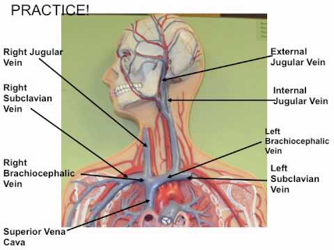
Coronary Vessels Anatomical Health Care Vector Illustration Labeled Diagram Heart Blood Flow System With Blood Vessel Scheme Stock Vector Illustration Of Heartbeat Cardio 116076791 from thumbs.dreamstime.com Blood vessels are the channels or conduits through which blood is distributed to body tissues. The vessels of the arms are part of the circulatory system, which provides nutrients to the tissues. Liver anatomy blood supply 19 photos of the liver anatomy blood supply anatomical location of liver, blood vessels that carry blood to the liver, dual blood supply to liver, functional anatomy of liver, liver and its functions, liver on the human body, normal anatomy of the liver, position of liver, inner body, anatomical location of … The smallest veins are called venules. This video covers one of our blood vessel models 4 but is clearly visible entering the right atrium of the heart. Blood is supplied to parts within the neck, head and brain through branches of the subclavian and common carotid arteries. Learn vocabulary, terms, and more with flashcards, games, and other study tools.
The iliac, femoral, popliteal and tibial (calf) veins are the deep veins in the legs.
Blood vessels of the abdomen and pelvis. Other sets by this creator. Blood vessels prepared by dr. Eventually, the smallest arteries, vessels called arterioles, further branch into tiny capillaries, where nutrients and wastes are exchanged. Disclosure • the material and the illustrations are adopted from the textbook human anatomy and physiology / ninth edition/ This article lists a series of labeled imaging anatomy cases by system and modality. Navigate to the cardiovascular system area in the following pal 3.0 module: Hma practical 3 for monday july 23 and wednesday july 25. Like arteries, veins form a complex, branching system of larger and smaller vessels. Blood vessel labeling 9p image quiz. Use key choices to identify the blood vessel tunic described. It extends on each side of the neck and divides at the level of the larynx into two branches: Blood vessels labeled diagram, blood vessels labeling exercises, cat blood vessels labeled, human anatomy blood vessels, human.
Blood is circulated through the body by blood vessels via the cardiovascular system which is comprised of the heart and the circulatory system.arteries move blood from the heart first to smaller arterioles, then capillaries or sinusoids, venules, veins, and back to the heart. Liver anatomy blood supply 19 photos of the liver anatomy blood supply anatomical location of liver, blood vessels that carry blood to the liver, dual blood supply to liver, functional anatomy of liver, liver and its functions, liver on the human body, normal anatomy of the liver, position of liver, inner body, anatomical location of … Start studying blood vessels labeling. Like arteries, veins form a complex, branching system of larger and smaller vessels. Human cadaver, anatomical models, histology, cat, and fetal pig.
Vesselsofabdomenandthorax from faculty.etsu.edu Veins return blood back toward the heart. Anatomy of blood vessels review sheet 32 261 microscopic structure of the blood vessels 1. Veins usually colored blue because oxygen poor, carry blood to the heart from the capillaries. Blood vessel labeling 7p image quiz. The smallest veins are called venules. Like arteries, veins form a complex, branching system of larger and smaller vessels. Capillaries come together to form venules, small blood vessels that carry blood to a vein, a larger blood vessel that returns blood to the heart. Elastic arteries (conducting arteries) are the largest arteries and include the aorta and other nearby branches.
Blood vessels 11p image quiz.
Human cadaver, anatomical models, histology, cat, and fetal pig. The vessels make up two closed systems of tubes that begin and end at the heart.one system, the pulmonary vessels, transports blood from the right ventricle to the lungs and back to the left atrium.the other system, the systemic vessels, carries blood from. Blood vessels of the abdomen and pelvis. The iliac, femoral, popliteal and tibial (calf) veins are the deep veins in the legs. Its smooth surface decreases resistance to blood flow The vessels that carry blood away from the heart are called arteries, and their very small branches are arterioles. Classification & structure of blood vessels. The word vascular, meaning relating to the blood vessels, is derived from the latin vas, meaning vessel. Veins usually colored blue because oxygen poor, carry blood to the heart from the capillaries. The smallest veins are called venules. Bulky middle tunic contains smooth muscle and elastin 3. That being said, all arterial blood delivered to this region comes via branches of the abdominal aorta, and all venous blood eventually finds its way back to. As the abdomen and pelvis contain the majority of internal organs, these regions need to be supplied by an extensive network of arteries and veins.
Arteries transport blood away from the heart. As the abdomen and pelvis contain the majority of internal organs, these regions need to be supplied by an extensive network of arteries and veins. Blood vessel labeling 9p image quiz. Disclosure • the material and the illustrations are adopted from the textbook human anatomy and physiology / ninth edition/ Other sets by this creator.
Blood Vessel Man Model Youtube from i.ytimg.com Use key choices to identify the blood vessel tunic described. Blood is supplied to parts within the neck, head and brain through branches of the subclavian and common carotid arteries. The inner lining is the endothelium and is. The three major types of blood vessels: The iliac, femoral, popliteal and tibial (calf) veins are the deep veins in the legs. Blood vessels are the specially designed tubes that carry blood throughout the body. Liver anatomy blood supply 19 photos of the liver anatomy blood supply anatomical location of liver, blood vessels that carry blood to the liver, dual blood supply to liver, functional anatomy of liver, liver and its functions, liver on the human body, normal anatomy of the liver, position of liver, inner body, anatomical location of … The common cartoid artery extends from the brachiocephalic artery.
The arteries deliver freshly oxygenated blood to muscles and bone.
The iliac, femoral, popliteal and tibial (calf) veins are the deep veins in the legs. 4 but is clearly visible entering the right atrium of the heart. Its smooth surface decreases resistance to blood flow That being said, all arterial blood delivered to this region comes via branches of the abdominal aorta, and all venous blood eventually finds its way back to. Blood vessel structure and function. Blood is supplied to parts within the neck, head and brain through branches of the subclavian and common carotid arteries. Arteries carry blood away from the heart. Human cadaver, anatomical models, histology, cat, and fetal pig. Veins (in blue) are the blood vessels that return blood to the heart. Very small branches that collect the blood from the various organs and parts are called venules, and they unite to form veins, which return the blood to the heart. Blood vessels 11p image quiz. This video covers one of our blood vessel models Blood vessels are the channels or conduits through which blood is distributed to body tissues.